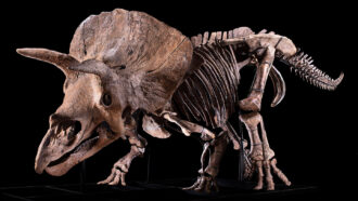
A gaping hole in the bony frill of a Triceratops dubbed “Big John” may be a battle scar from one of his peers.
The frill that haloes the head of Triceratops is an iconic part of its look. Equally iconic, at least to paleontologists, are the holes that mar the headgear. For over a century, researchers have debated various explanations for the holes, called fenestrae — from battle scars to natural aging processes. Now, a microscopic analysis of Big John’s partially healed lesion suggests that it could be a traumatic injury from a fight with another Triceratops, researchers report April 7 in Scientific Reports.
In summer 2021, Flavio Bacchia, director of Zoic LLC in Trieste, Italy, was reconstructing the skeleton of Big John, the largest known Triceratops to date, when he noticed a keyhole-shaped fenestra on the right side of its frill. Bacchia then reached out to Ruggero D’Anastasio, a paleopathologist at the “G. D’Annunzio” University of Chieti-Pescara in Italy who studies injuries and diseases in ancient human and other animal remains.
“When I saw, for the first time, the opening, I realized that there was something strange,” D’Anastasio says. In particular, the irregular margins of the hole were odd. He had never seen anything like it.
To analyze the fossilized tissues around the fenestra, he obtained a piece of bone about the size of a 9-volt battery, cut from the bottom of the keyhole. The rest of Big John sold at an auction for $7.7 million — the most expensive non–Tyrannosaurus rex dinosaur fossil ever.
Looking at the bone under a scanning electron microscope, D’Anastasio and his team found evidence consistent with the formation processes of new bone that are usually observed in mammals. New bone growth is typically supported by blood vessels, and in the bone near the border of the hole, the tissue was porous and strewn with vascular canals. Farther from the fenestra, the bone showed little evidence of the vessels.
The team found that the irregularity of the hole margins that D’Anastasio had observed was also present at the microscopic level. The border was dappled with microscopic dimples called Howship lacunae, where, in one of the first steps of bone healing, bone cells eroded the existing bone to be replaced with healthy bone. The researchers also observed primary osteons, formations that occur during new bone growth.
In addition, a chemical analysis revealed high levels of sulfur, indicative of proteins involved in new bone formation. In mature bones, sulfur is present in only low quantities.
Taken all together, it was clear that this particular fenestra was a partially healed wound. “The presence of healing bone is typical of the response to a traumatic event,” D’Anastasio says.
Scientists can only hypothesize what happened so long ago. But the location and shape of the wound suggest that Big John’s frill was impaled from behind by a Triceratops rival, adding evidence to the idea that Triceratops fought with one another (SN: 1/27/09). It was probably an initial puncture that was pulled downward to create the keyhole shape, the researchers say.
“Pathology is a great tool to understand the behavior of dinosaurs,” says Filippo Bertozzo, a dinosaur paleontologist at the Royal Belgian Institute of Natural Sciences in Brussels who was not involved in the study. Dinosaur behavior has long been in the realm of speculation, he says, but analyses like these can provide a glimpse into the lifestyle of these animals.
He adds that this particular wound is “not a Rosetta stone,” because it’s unlikely that all fenestrae are battle injuries. “Fenestration is still a big mystery.”
What’s also a mystery, D’Anastasio says, is why the bone remodeling seen in this Triceratops sample was more similar to healing observed in mammals than in other dinosaurs. And Big John himself might hold more secrets.
“We published an aspect, a paleopathological case,” D’Anastasio says. “The complete skeleton of Big John must be studied.”

 A new treatment could restore some mobility in people paralyzed by strokes
  What has Perseverance found in two years on Mars?
  This robot automatically tucks its limbs to squeeze through spaces
  Greta Thunberg’s new book urges the world to take climate action now
  Glassy eyes may help young crustaceans hide from predators in plain sight
  A chemical imbalance doesn’t explain depression. So what does?